What is the Difference Between a Vision Test and an Eye Exam?
Many people think that an eye exam is simply a test to see if you have any change of vision but this isn’t actually the case. A vision test checks your eyes for any changes in vision but does not detect the underlying cause of those vision changes or any eye damage or disease that may be present.
An eye exam , on the other hand is a detailed examination of the eye, looking at the structure and health of the eye as well as all aspects of vision via a number of specialised machines or tools . A vision test may be included in a comprehensive eye exam, but in many cases a vision test may lead an optometrist to perform a comprehensive eye exam.
How Often Should I Have a Comprehensive Eye Exam?
How often you should have your eyes tested depends on a number of factors including age, overall health and any underlying risk factors that may increase your chance of developing eye problems. As a baseline, it’s recommended in Australia that people get their eyes tested annually so any changes in vision or developing eye problems may be picked up and treated as soon as possible.
People living with diabetes or already diagnosed with a degenerative eye disease may need to have their eyes tested more often if their optometrist/ophthalmologist advises. People with a family history of vision problems of loss, those taking medicines with potential vision side effects and heavy smokers may also require more frequent testing,
Do I Have to Pay For an Eye Exam?
In Australia, Medicare may either fully or partially cover the cost of an eye exam. The medicare rebate for exams is $57.70, and many optometry practices don’t charge any extra fee, meaning that you can get the test for free. If your optometrist doesn’t bulk bill you’ll have to pay for the exam first, then collect the rebate from Medicare. Many practices allow you to do this part electronically straight after your eye exam.
Anyone under the age of 65 with a valid and current Medicare card is eligible for one rebate for an eye exam every three years, unless new symptoms arise. In the case of new symptoms, such as blurred vision and vision headaches, new eye exams will be covered by Medicare as needed.
Similarly, people with pre-existing conditions or risk factors for eye damage, such as glaucoma, retinopathy and diabetes are eligible to have Medicare covered eye exams as often as needed.
How Are My Eyes Tested?
Your eye test will be carried out by either an optometrist or an ophthalmologist. While both of these eye specialists are equally able to perform eye exams, ophthalmologists and optometrists have different skill sets overall.
- Optometrist - an optometrist is specialised in testing, diagnosing and treating vision problems and prescribing corrective lenses (glasses and contact lenses). An optometrist may also be able to prescribe medications to treat eye ailments such as conjunctivitis or a stye.While an optometrist may diagnose serious eye problems such as retinopathy, glaucoma or cataracts, treatment may require a referral to an ophthalmologist. You do not require a referral to see an optometrist but your GP may recommend you see one if you are experiencing vision related problems
- Ophthalmologist - an opthamologist is a medical doctor that, in addition to providing comprehensive eye exams and prescribing corrective lenses, are also trained in diagnosing and treating complex eye problems and administering treatment. This treatment may include surgery.
Ophthalmologists may also be licensed to perform some corrective surgeries, such as laser eye surgery used to correct some cases of long or short-sightedness. A referral from a GP, optometrist or other specialist is required for an initial appointment with an ophthalmologist.
The exact tests performed may vary a little between different optometrists or if a certain problem is being searched for, but a comprehensive eye exam typically consists first of collecting patient history. If there are any particular risk factors or genetic predispositions, the optometrist may opt for additional tests or focus on one group of tests.
The fastest and easiest way to book an eye test is to search and book online with MyHealth1st.
If you have an existing prescription, bringing your existing glasses gives the optometrist a baseline to work off to see if there has been any change in vision.If you normally wear contact lenses, ask your optometrist whether he or she would prefer you to attend your eye exam with or without wearing your contact lenses.
Tests that may be part of a comprehensive eye exam include:
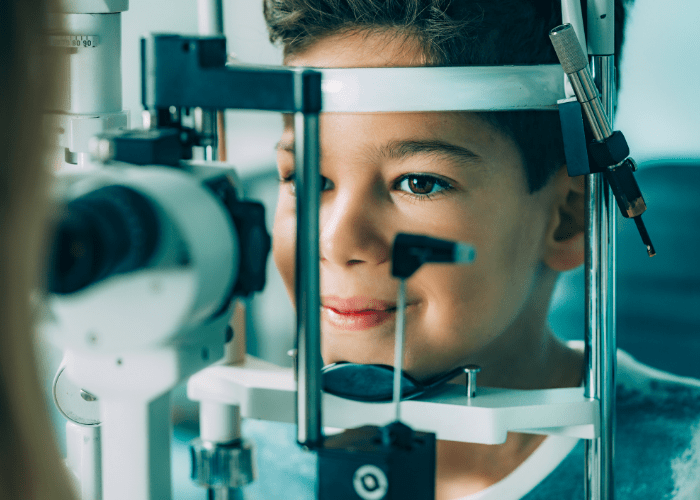
- Slit Lamp Exam - a slit lamp is a kind of specialised microscope that allows an optometrist to evaluate the health of your eyes by giving them a magnified look at the structures of your eyes. This enables them to erect any amage, infection or disease that may be present. During a slit lamp test, your optometrist will have you place your chin in a chin rest and a light will be shone into your eye. This light is highly variable, and using it, the optometrist is able to examine both surface and underlying structures in the eye. First they will examine the outside of the eye - cornea, eyelids, conjunctiva, iris and the like - and then move on to deeper structures such as the macula, retina and optic nerve. The slit lamp allows for the diagnosis of a number of conditions, such as macular degeneration, diabetic retinopathy and cataracts.
- Tonometry (Puff Test) - the puff test is one of the primary ways in which glaucoma is diagnosed. During a puff test, the patient places their chin in the chin rest of a machine. A harmless and painless puff of air is directed into the eye, measuring eye pressure. The tonometer measures the resistance the eye has to the puff and from that measures the intraocular pressure (IOP), or pressure in the eye. High IOP is both a risk factor for and possible indicator of glaucoma. There are other equally accurate ways of measuring the pressure of your eyes. These can involve using eye drops, but these drops do not affect your vision in any way.
- Ocular Cohesion Tomography (OCT) - the most advanced form of optical imaging currently available, Ocular Cohesion Tomography creates 3D images of the retina in-situ by compiling cross-sectional images of the retina. This allows the eye care professional to look at the retina as a whole or to look at individual cross sections within the retina to diagnose a range of conditions and diseases. OCT may be used to check for retinal diseases such as diabetic retinopathy, macular degeneration and glaucoma. Due to the detail of the image, OCT may also be a valuable way of documenting and monitoring any changes in the eye.
- Digital Retinal Photography - this specialised camera can generate high quality diagnostic images which can be used to diagnose disease either straight away, or using them as a baseline to compare with any future changes.
- Ocular Motility - each eye has six muscles that control its movement through contracting and relaxing, each often in cooperation with other muscles. These muscles move the eye up, down and to the sides. An ocular motility test checks to see if these muscles are working correctly. The patient is asked to follow a light or object with their eyes, typically in a “H” pattern, and the optometrist monitors the eyes, examining the speed, smoothness, range and symmetry of the movement. Inaccurate, jerky or otherwise anomalous movement may be an indicator of a number of conditions such as strabismus (misaligned eyes), amblyopia (lazy eye), extraocular muscle dysfunction or potential nerve damage/palsy caused by damage, disease (such as diabetes or multiple sclerosis), stroke, vitamin deficiency or alcohol intake.
- Pupillary Dilation - dilation of the pupils allows the optometrist to get a good look at the inside of your eyes. Drops that dilate the eye are placed in each eye. These may take 15-30 minutes to take full effect. Once the pupils are fully dilated the optometrist uses various lights and scopes to look into your eyes, examining the internal structure. While dilation isn’t painful, you may experience sensitivity to light and difficulty focussing on things close to you while your pupils are dilated. It may take a few hours for your eyes to return to normal after dilation, so you should bring a pair of sunglasses if you are having a dilation to protect your eyes and make yourself more comfortable afterwards. You should not drive after having a dilation test.
- Visual Acuity Test - the visual acuity test is the most recognisable of eye tests. Patients are asked to read from a Snellen chart, a pyramidal chart of a number of lines of letters, with the size of the letters becoming smaller the further down the chart they are. The patient reads as far down the chart as possible, first with a single eye (covering the other) and then with both eyes to judge their distance vision. A smaller chart is used to judge close vision in a similar manner. Another test, known as the Random E, may be used to test visual acuity as well. In this test, the patient is asked to identify in which direction the letter E is facing - up, down, left, or right. Visual acuity tests are used to assess distance (both far and near) vision and may diagnose disorders such as myopia (short-sightedness) and hyperopia (far-sightedness).
- Refractive Error Test - this test is used to see if there are any problems in the way that your eyes focus light onto your retina. This is either done manually or with a machine that measures the amount of light reflected by your retina. The more accurate the optometrist is with this test, usually the better your glasses or contact lenses will make you see! A refractive error test is useful in diagnosing astigmatism (refractive problems related to the shape of the lens), myopia, hyperopia, presbyopia (an age-related condition of the lens) and may be helpful in diagnosing macular degeneration, retinal vessel occlusion, retinal detachment and retinitis pigmentosa.
- Visual Field Test - this test checks peripheral vision. During the test, one eye is tested at a time, with the other being covered. There are a number of different tests that may be used, such as the Amsler Grid, Humphrey Field Analyser or Goldmann Perimeter test, but all require the eye to be kept still while focussing on one point. Depending on the test used, patients may have to press a button when something appears a light appears in the periphery of their vision, whether a grid appears solid, faded, distorted or missing, or otherwise indicate things appearing or disappearing in peripheral vision. Visual field testing may be helpful in screening for glaucoma or ptosis (lid droop), hydroxychloroquine toxicity or optic nerve conditions (including tumours).
- Colour Blindness Test - more accurately known as a Colour Vision Deficiency test, these tests check to see if visual problems may be due to an inability to properly differentiate some colours. The most effective and best known colour vision deficiency test is known as the Ishihara test. In this test, people are presented with a number of images showing numbers consisting of a number of coloured dots on a field of other dots of another colour (red/green, blue/yellow, etc). If you wear glasses or contact lenses, or have been diagnosed with a condition that may affect your eyes, you should have an annual eye test to monitor the condition of your eyes. Even if your eyes are healthy, you should still have an eye test every few years to make sure that nothing has changed.